Perfusion CT – An Interview with Dr. Emily Clewett
CT perfusion, along with CT angiography (CTA), can be a very important adjunct when treating strokes as opposed to conventional unenhanced CT brain imaging. Emily Clewett, Head of Section of Emergency Radiology Scandinavia, and the emergency senior clinical advisor for the AI Centre of Excellence has led her team to include CT perfusion in the routine workout of stroke patients for selected client hospitals in Sweden. Emily has been working on this project for a year now, so it is about time to ask her some questions about it:
What is Perfusion CT and why is it important in stroke diagnosis?
EC: In a stroke setting, it is vital to provide treatment as soon as possible – ‘time is brain’. We use several methods to find the cause of the patients’ symptoms, where the plain (non-contrast) CT head allows us to spot bleeds or established infarcts and the CT angiogram depicts the site of occlusion (blood clot).
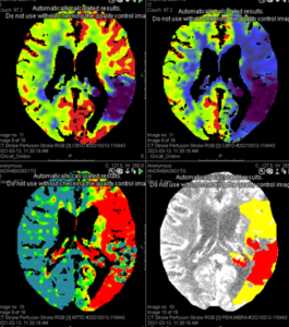
The next level in stroke diagnostics is the CT perfusion, adding information about how much of the brain is at risk if the clot isn´t removed and helping the neuro-interventionists with selecting the patients who would best benefit from an endovascular procedure for the mechanic removal of the thrombus (some patients are treated only medically, with thrombus-dissolving medications). The perfusion scan is obtained after injection of intravenous contrast agent; the CT scanner acquires images of the same area of the brain at different timepoints, measuring the inflow of contrast agent and thus adding a temporal dimension.

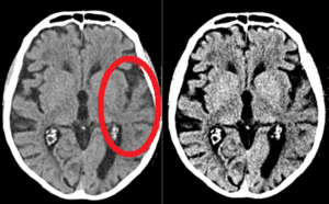
Acute infarct on plain CT head

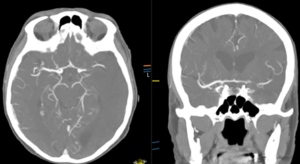
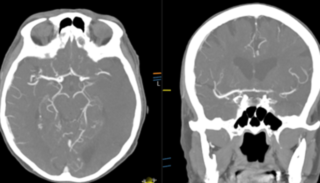
Occlusion (blood clot) on CT Angiogram

Perfusion scan showing established infarct (red, bottom right) vs area, which is ischaemic, but not yet infarcted, and thus salvable (yellow, bottom right)
Which role do Artificial Intelligence (AI) and post-processing software play in the interpretation of CT perfusion?
EC: The Emergency section has been an early adopter of AI-based diagnostic tools for the interpretation of CT head. A stroke with a bleed requires very different treatment from one with a blood clot, and it is vital that the radiologist identifies even the smallest of bleedings At Telemedicine Clinic, AI solutions help in ensuring this is done correctly
The images from a CT perfusion scan must be post-processed; this can be done manually or using special software with an automated workflow. There are AI software capable of processing perfusion scans, an emerging and exciting field in Sweden right now!
What challenges did you encounter when introducing this new diagnostic service?
EC: While CT perfusion imaging has been around for quite some time, it is usually reported by highly specialized neuroradiologists. We have worked intensively to train our radiologist in this particular method. With the help of our Swedish client Örebro University Hospital which is pioneering the stroke care / teleradiology synergy, we created a portfolio of anonymised practice cases complete with reports and tutorials. To guarantee the highest level of quality and safety, we use prospective double reading for all stroke pathway cases, including those with CT perfusion images, which means all emergency reports are signed by two radiologists before being distributed.
Do you see this as a service that will become more popular and offered to more client hospitals?
EC: A couple of years ago new national guidelines were presented in Sweden, suggesting more stroke patients can benefit from endovascular mechanical clot removal. Perfusion scans are vital for patient selection, and we are currently seeing more frequent requests for this service. It is our goal to eventually be able to offer perfusion reporting to all our clients.


